Ejection Fraction

During my annual cardiac check-up with my cardiologist on Monday, the nurse performed an ultrasound scan (sonogram) of my heart. An ultrasound is a non-invasive procedure that uses sound waves, which bounce off solid structures to create two-dimensional black-and-white images on a screen.
This test helps cardiologists diagnose a range of heart conditions. It’s commonly used to assess blood flow through the heart and its valves. The results can help identify heart disease and measure important indicators such as the ejection fraction (EF).
When someone experiences a heart attack, the heart muscle can suffer damage depending on how quickly treatment is received. An ultrasound can detect this damage and measure how effectively the heart is pumping—this is known as the ejection fraction.
In my case, I’ve been fortunate. Both of my heart attacks were treated promptly with emergency surgery to insert stents into the blocked arteries, restoring blood flow to the heart muscle.
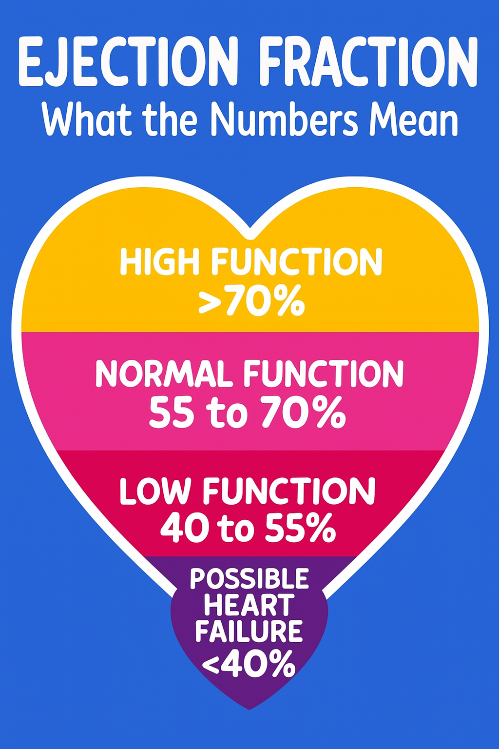
During my recent scan, my ejection fraction was measured at 64%, which falls within the healthy range. However, the scan also revealed Grade 1 diastolic dysfunction with normal filling pressure.
This was the first time I truly paid attention to the concept of ejection fraction, and I became curious about what the percentage levels mean for heart patients and healthy adults my age (62).
So I asked my cardiologist, “What is a normal ejection fraction?” He referred me to a table commonly used by heart health professionals to classify ejection fraction ranges—see below:
Grade 1 diastolic dysfunction is commonly referred to as "impaired relaxation." In patients with this grade of dysfunction, the diastolic filling of the ventricles is slower than normal, but other cardiac measurements typically remain within normal limits.
Healthcare providers use a grading system to assess the severity of diastolic dysfunction:
-
Grade I: Slightly impaired diastolic function. This is a common finding in individuals over the age of 60.
-
Grade II: Indicates elevated pressure in the left side of the heart.
-
Grade III: Reflects significantly elevated pressure in the left side of the heart.
During the procedure, I was also connected to an ECG (electrocardiogram) monitor so the healthcare provider could assess my heart’s electrical activity. Thankfully, the ECG results were normal. An ECG is a simple, non-invasive test that records the heart’s electrical signals and helps diagnose conditions such as abnormal heart rhythms and coronary heart disease, including heart attacks and angina.
Overall, my annual health check went well.


